
Project 3369: B. M. Gee, J. J. Bevitt, R. R. Reisz, M. Ruta. 2020. Computed tomographic analysis of the cranium of the early Permian recumbirostran ‘microsaur’ Euryodus dalyae reveals new details of the braincase and mandible. Papers in Palaeontology:null.
Abstract
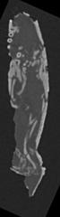
Recumbirostran ‘microsaurs’ are a clade of Palaeozoic tetrapods that possess numerous morphological adaptations for fossorial ecologies. Re-study of many ‘microsaurs’ using tomographic methods has provided substantial new data on poorly known anatomy that informs their debated phylogenetic position. Recent studies have identified suites of features among recumbirostrans that place the group within crown Amniota, contrary to hypothesized positions on the amniote stem or the lissamphibian stem. Herein we describe the cranial anatomy of the early Permian gymnarthrid Euryodus dalyae through tomographic analysis of the holotype from South Grandfield, Oklahoma and new specimens from karst deposits near Richards Spur. The braincase of E. dalyae is composed of well-ossified pleurosphenoids, orbitosphenoids that brace against the skull roof, and unpaired median ossifications. The otic capsules are well-ossified, and the occiput is unconsolidated. Analysis of the mandibles, typically obscured in articulated specimens, reveals a second tooth row on the dentary, a feature previously unknown in ‘microsaurs’ that is reminiscent of the condition of the co-occurring captorhinid Captorhinus aguti. The Richards Spur specimens share many of these features, including the second tooth row, but the neurocranium of the scanned specimen (OMNH 53519) differs from that of the holotype of E. dalyae (e.g. absence of unpaired median ossifications), and these specimens are referred to Euryodus sp. These data add to the growing neurocranial dataset of ‘microsaurs’, which is essential for iterative reevaluation of early tetrapod phylogeny. This discovery of multiple tooth rows in ‘microsaurs’ provides further support for the hypothesized close relationships between ‘microsaurs’ and reptiles.Read the article »
Article DOI: 10.1002/spp2.1304
Project DOI: 10.7934/P3369, http://dx.doi.org/10.7934/P3369
| This project contains |
|---|
Download Project SDD File |
Currently Viewing:
MorphoBank Project 3369
MorphoBank Project 3369
- Creation Date:
23 January 2019 - Publication Date:
13 March 2020 - Project views: 16760

- Media downloads: 16
Authors' Institutions ![]()
- Jilin University
- University of Toronto
- Australian Nuclear Science and Technology Organisation
Members
| member name | taxa |
specimens |
media |
| Bryan Gee Project Administrator | 2 | 2 | 15 |
Project has no matrices defined.
Project views
| type | number of views | Individual items viewed (where applicable) |
| Total project views | 16760 | |
| Project overview | 1428 | |
| Media views | 7630 | Media search (2319 views); M685189 (360 views); M685190 (339 views); M685193 (371 views); M685188 (356 views); M685191 (337 views); M685194 (348 views); M685195 (327 views); M685196 (398 views); M685238 (376 views); M685239 (368 views); M685240 (353 views); M685242 (353 views); M685243 (352 views); M685244 (349 views); M685245 (324 views); |
| Views for media list | 2693 | |
| Folio views | 1266 | Folio list (557 views); 16-bit TIFF stack for OMNH 53519 (349 views); 16-bit TIFF stack for FM UR 2296 (360 views); |
| Specimen list | 1661 | |
| Taxon list | 877 | |
| Bibliography | 532 | |
| Documents list | 673 |
Project downloads
| type | number of downloads | Individual items downloaded (where applicable) |
| Total downloads from project | 196 | |
| Media downloads | 16 | M685189 (3 downloads); M685194 (2 downloads); M685196 (4 downloads); M685193 (3 downloads); M685195 (1 download); M685188 (2 downloads); M685190 (1 download); |
| Document downloads | 21 | Modified character matrix of Pardo et al. (2017, Nature) (11 downloads); README (pdf) for FMNH UR 2296 scan (1 download); README (text file) for OMNH 53519 (1 download); Supplemental figures (8 downloads); |
| Project downloads | 159 |